 |
|
|
| Home
Special Opportunities for Members Website developed by
|
Items of Interest Risk Management Issues with EHR Efficiency Tools Many of the software programs used for electronic records include time saving features such as “copy forward,” bringing the last note to the current visit, prewritten “standard” procedure notes and prepopulated text when a box is checked. While these tools save time in a busy patient day, they should be used with care. Records that have identical entries can raise the question of whether the care was actually performed. •
When copying and pasting information, always review and make changes based on the
patient's comments today. Only copy and paste information relevant to today’s visit. Problems from a previous visit that are resolved should not be in the chief complaint of the next visit. In addition to the issues created in defending malpractice claims, Medicare is also auditing for cloning of records as potential fraud. The work value and payment is based on performing the elements of the history and examination. They have developed software that can identify blocks of identical text in patient records. On audit they can deny the claim, requiring refund of payments and possible fines. Review the chart note that prints out and is sent for an audit to make sure all the relevant documentation is included. Down coding can occur based on missing exam or history elements that don’t appear in the printed chart note even though they are documented in the record. Volunteers Needed for Blind Sports Organization
(BSO) E-Learning for eye care professionals made quick and easy. Tips for
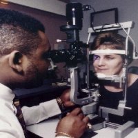
Taking Fundus Photos 1) Communicate with your patient! 2) If you see something of
interest - photograph it! Document everything! 3) Let your patients know that they can blink as needed, BUT to open eyes wide between blinks. If you don’t tell them they can blink, they will try not to. This will cause stress on the patient, strain, squinting, tearing, and poor image quality. Let your patients blink! 4) Use the fixation pointer if the patient has a difficult time seeing the fixation light. However, if the patient is cooperative, since the pointer may distract from the photograph, pull it out of the lens at the last moment before clicking the shutter. 5) For light sensitive patients, turn the viewing light down low. Try to line up the eye as best you can peeking through their squinted eyes. You should be able to line up their vessels, find their optic nerve, etc. Try to obtain the best focus possible like this. Once you have everything lined up, have the patient blink, and then lift their lid with a Q Tip and shoot. You may need to make a few adjustments, but you’ll definitely get a better photo than if you held their eye up through the entire process. 6) When taking stereo optic nerve photographs, focus the first image on the rim of the optic nerve head and the second image on the cup. This will give the pair a much more appreciative three dimensional view. 7) When shooting images for a montage, photograph the fields in a clockwise or counter-clockwise direction beginning always with the posterior pole. This will help you to make sure you have obtained all necessary fields and the order helps the computer software match up the images more accurately. 8) If you’re having a difficult time getting a clear view of the eye, pull the camera back a bit and set the focus lens on A. This will give you a view of the opacity and how to get around it to obtain a clearer fundus image. And while you’re at it, take a photograph of the opacity. Document everything! Links Online Portal for Ophthalmic Technicians Wills Eye Hospital® Annual Alumni Conference Technicians' Program American Academy of Ophthalmology American Society of Cataract & Refractive Surgery and American Society of Ophthalmic Administrators Macular Degeneration Foundation American Printing House for the Blind Centers
for Medicare & Medicaid Services (CMS) American Society of Ophthalmic Administrators Associated Services for the Blind Our thanks to Diane Brash of the Associated Services for the Blind. Diane has provided us with resources to assist our visually impaired patients:Associated
Services for the Blind Library
for the Blind, GED programs: PA
Bureau of Blindness and Visual Services: PA
Association for the Blind:
|
|
|
PROS MESSAGE CENTER 215-825-4725 Email: mmassini@willseye.org |